By continuing to use the website or clicking Accept you consent to our cookies and personal data policy and confirm that you are at least 18 year old. For details please see Privacy Policy and Terms
Accept
Likes
Comments
Share

@smoking_hills9
Follow
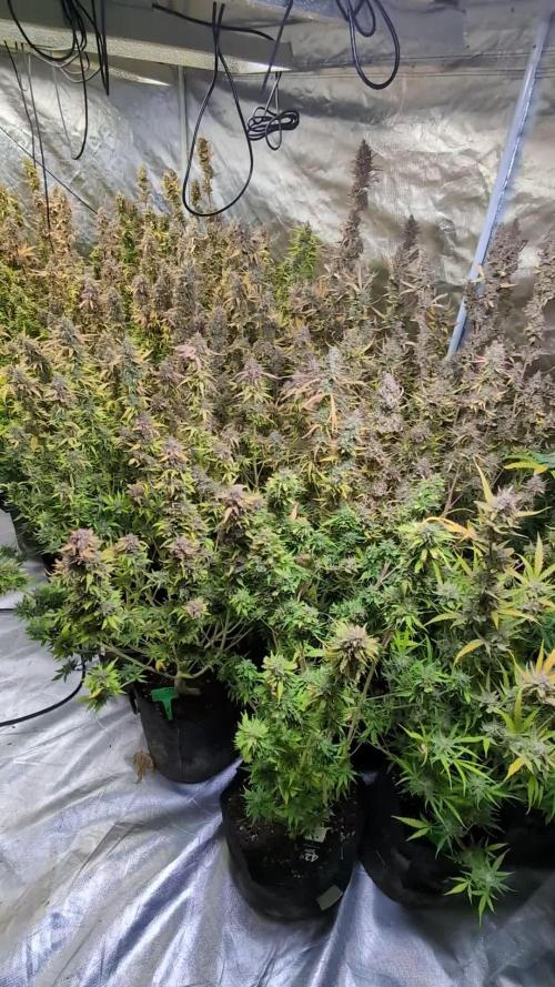
Some beautiful plants especially 1 and 2. Sweet skittles smell. Happy with outcome.

Likes
2
Share


@LadyGreen
Follow
She growing well, so well in fact I have started to HST the off shoot branches to keep the grow low. They are nice and green still so easy to HST and take to it well.
It's getting ready to flip but waiting on my new tent...so she will just have to keep growing taking over my veg cabinet

Likes
18
Share


@Mazgoth
Follow
Not really big buds but going really good,they have time to grow more flowers but don't hope too much,still love them,D53(Update)I purchase an CFL maxibright 200W and I'm gonna still with that until harvest

Likes
21
Share


@cal917
Follow
Noticed the blueberry requires lots of nitrogen and stretches much. All plants fed nutrients 1 per week and microbes once per week with regular watering in between. Start of 5th week of flower.


Likes
12
Share


@Nxs235
Follow
Hello community, today is day 114 from dry sunflower seeds. Last week before harvest, watering with bottled water all week. The smell of ripe melon, fruity and tart.
Likes
11
Share


@Guerilla_Jack
Follow
Hello growers an breeders.this is the end of flowering time for my girls.i flash them with flawless finish this week.smoke report in week🙂


Likes
6
Share


@Beanz2Budz
Follow
Tip - Before every watering I ensure to leave the water rest for a minimum of 24-36hours to allow for chlorine dissipation.

Likes
25
Share


@Ultraviolet
Follow
ANTHOCYANIN production is primarily controlled by the Cryptochrome (CR1) Photoreceptor ( !! UV and Blue Spectrums are primary drivers in the production of the pigment that replaces chlorophyll, isn't that awesome!
1. Diverse photoreceptors in plants
Many civilizations, including the sun god of ancient Egypt, thought that the blessings of sunlight were the source of life. In fact, the survival of all life, including humans, is supported by the photosynthesis of plants that capture solar energy. Plants that perform photosynthesis have no means of transportation except for some algae. Therefore, it is necessary to monitor various changes in the external environment and respond appropriately to the place to survive. Among various environmental information, light is especially important information for plants that perform photosynthesis.
In the process of evolution, plants acquired phytochrome, which mainly receives light in the red light region, and multiple blue light receptors, including his hytropin and phototropin, in order to sense the light environment. .. In addition to these, an ultraviolet light receptor named UVR8 was recently discovered. The latest image of the molecular structure and function of these various plant photoreceptors (Fig. 1), focusing on phytochrome and phototropin.
Figure 1
Ultraviolet-visible absorption spectra of phytochrome, cryptochrome, phototropin, and UVR8. The dashed line represents each bioactive absorption spectrum.
2. Phytochrome; red-far red photoreversible molecular switch
What is phytochrome?
Phytochrome is a photochromic photoreceptor, and has two absorption types, a red light absorption type Pr (absorption maximum wavelength of about 665 nm) and a far-red light absorption type Pfr (730 nm). Reversible light conversion between the two by red light and far-red light, respectively(Fig. 1A, solid line and broken line). In general, Pfr is the active form that causes a physiological response. With some exceptions, phytochrome can be said to function as a photoreversible molecular switch. The background of the discovery is as follows. There are some types of plants that require light for germination (light seed germination). From that study, it was found that germination was induced by red light, the effect was inhibited by subsequent far-red light irradiation, and this could be repeated, and the existence of photoreceptors that reversibly photoconvert was predicted. In 1959, its existence was confirmed by the absorption spectrum measurement of the yellow sprout tissue, and it was named phytochrome. Why does the plant have a sensor to distinguish between such red light and far-red light? There is no big difference between the red and far-red light regions in the open-field spectrum of sunlight, but the proportion of red light is greatly reduced due to the absorption of chloroplasts in the shade of plants. Similar changes in light quality occur in the evening sunlight. Plants perceive this difference in light quality as the ratio of Pr and Pfr, recognize the light environment, and respond to it.
Subsequent studies have revealed that it is responsible for various photomorphogenic reactions such as photoperiodic flowering induction, shade repellent, and deyellowing (greening). Furthermore, with the introduction of the model plant Arabidopsis thaliana (At) and the development of molecular biological analysis methods, research has progressed dramatically, and his five types of phytochromes (phyA-E) are present in Arabidopsis thaliana. all right. With the progress of the genome project, Fi’s tochrome-like photoreceptors were found in cyanobacteria, a photosynthetic prokaryotes other than plants. Furthermore, in non-photosynthetic bacteria, a homologue molecule called bacteriophytochrome photoreceptor (BphP) was found in Pseudomonas aeruginosa (Pa) and radiation-resistant bacteria (Deinococcus radiodurans, Dr).
Domain structure of phytochrome molecule
Phytochrome molecule can be roughly divided into N-terminal side and C-terminal side region. PAS (Per / Arndt / Sim: blue), GAF (cGMP phosphodiesterase / adenylyl cyclase / FhlA: green), PHY (phyto-chrome: purple) 3 in the N-terminal region of plant phytochrome (Fig. 2A) There are two domains and an N-terminal extension region (NTE: dark blue), and phytochromobilin (PΦB), which is one of the ring-opening tetrapyrroles, is thioether-bonded to the system stored in GAF as a chromophore. ing. PAS is a domain involved in the interaction between signal transduction-related proteins, and PHY is a phytochrome-specific domain. There are two PASs and her histidine kinase-related (HKR) domain (red) in the C-terminal region, but the histidine essential for kinase activity is not conserved.
3. Phototropin; photosynthetic efficiency optimized blue light receptor
What is phototropin?
Charles Darwin, who is famous for his theory of evolution, wrote in his book “The power of move-ment in plants” published in 1882 that plants bend toward blue light. Approximately 100 years later, the protein nph1 (nonphoto-tropic hypocotyl 1) encoded by one of the causative genes of Arabidopsis mutants causing phototropic abnormalities was identified as a blue photoreceptor. Later, another isotype npl1 was found and renamed phototropin 1 (phot1) and 2 (phot2), respectively. In addition to phototropism, phototropin is damaged by chloroplast photolocalization (chloroplasts move through the epidermal cells of the leaves and gather on the cell surface under appropriate light intensity for photosynthesis. As a photoreceptor for reactions such as escaping to the side of cells under dangerous strong light) and stomata (reactions that open stomata to optimize the uptake of carbon dioxide, which is the rate-determining process of photosynthetic reactions). It became clear that it worked. In this way, phototropin can be said to be a blue light receptor responsible for optimizing photosynthetic efficiency.
Domain structure and LOV photoreaction of phototropin molecule
Phototropin molecule has two photoreceptive domains (LOV1 and LOV2) called LOV (Light-Oxygen-Voltage sensing) on the N-terminal side, and serine / on the C-terminal side. It is a protein kinase that forms threonine kinase (STK) (Fig. 4Aa) and whose activity is regulated by light. LOV is one molecule as a chromophore, he binds FMN (flavin mononucleotide) non-covalently. The LOV forms an α/βfold, and the FMN is located on a β-sheet consisting of five antiparallel β-strands (Fig. 4B). The FMN in the ground state LOV shows the absorption spectrum of a typical oxidized flavin protein with a triplet oscillation structure and an absorption maximum wavelength of 450 nm, and is called D450 (Fig. 1C and Fig. 4E). After being excited to the singlet excited state by blue light, the FMN shifts to the triplet excited state (L660t *) due to intersystem crossing, and then the C4 (Fig. 4C) of the isoaroxazine ring of the FMN is conserved in the vicinity. It forms a transient accretionary prism with the tain (red part in Fig. 4B Eα) (S390I). When this cysteine is replaced with alanine (C / A substitution), the addition reaction does not occur. The effect of adduct formation propagates to the protein moiety, causing kinase activation (S390II). After that, the formed cysteine-flavin adduct spontaneously dissociates and returns to the original D450 (Fig. 4E, dark regression reaction).
Phototropin kinase activity control mechanism by LOV2
Why does phototropin have two LOVs? Atphot1 was found as a protein that is rapidly autophosphorylated when irradiated with blue light.
The effect of the above C / A substitution on this self-phosphorylation reaction and phototropism was investigated, and LOV2 is the main photomolecular switch in both self-phosphorylation and phototropism. It turns out that it functions as. After that, from experiments using artificial substrates, STK has a constitutive activity, LOV2 functions as an inhibitory domain of this activity, and the inhibition is eliminated by photoreaction, while LOV1 is kinase light. It was shown to modify the photosensitivity of the activation reaction. In addition to this, LOV1 was found to act as a dimerization site from the crystal structure and his SAXS. What kind of molecular mechanism does LOV2 use to photoregulate kinase activity? The following two modules play important roles in this intramolecular signal transduction.
Figure 4
(A) Domain structure of LOV photoreceptors. a: Phototropin b: Neochrome c: FKF1 family protein d: Aureochrome (B) Crystal structure of auto barley phot1 LOV2. (C) Structure of FMN isoaroxazine ring. (D) Schematic diagram of the functional domain and module of Arabidopsis thaliana phot1. L, A’α, and Jα represent linker, A’α helix, and Jα helix, respectively. (E) LOV photoreaction. (F) Molecular structure model (mesh) of the LOV2-STK sample (black line) containing A’α of phot2 obtained based on SAXS under dark (top) and under bright (bottom). The yellow, red, and green space-filled models represent the crystal structures of LOV2-Jα, protein kinase A N-lobe, and C-robe, respectively, and black represents FMN. See the text for details.
1) Jα. LOV2 C of oat phot1-to α immediately after the terminus
Rix (Jα) is present (Fig. 4D), which interacts with the β-sheet (Fig. 4B) that forms the FMN-bound scaffold of LOV2 in the dark, but unfolds and dissociates from the β-sheet with photoreaction. It was shown by NMR that it does. According to the crystal structure of LOV2-Jα, this Jα is located on the back surface of the β sheet and mainly has a hydrophobic interaction. The formation of S390II causes twisting of the isoaroxazine ring and protonation of N5 (Fig. 4C).
As a result, the glutamine side chain present on his Iβ strand (Fig. 4B) in the β-sheet rotates to form a hydrogen bond with this protonated N5. Jα interacts with this his Iβ strand, and these changes are thought to cause the unfold-ing of Jα and dissociation from the β-sheet described above. Experiments such as amino acid substitution of Iβ strands revealed that kinases exhibit constitutive activity when this interaction is eliminated, and that Jα plays an important role in photoactivation of kinases.
2) A’α / Aβ gap.
Recently, several results have been reported showing the involvement of amino acids near the A’α helix (Fig. 4D) located upstream of the N-terminal of LOV2 in kinase photoactivation. Therefore, he investigated the role of this A’α and its neighboring amino acids in kinase photoactivation, photoreaction, and Jα structural change for Atphot1. The LOV2-STK polypeptide (Fig. 4D, underlined in black) was used as a photocontrollable kinase for kinase activity analysis. As a result, it was found that the photoactivation of the kinase was abolished when amino acid substitution was introduced into the A’α / Aβ gap between A’α and Aβ of the LOV2 core. Interestingly, he had no effect on the structural changes in Jα examined on the peptide map due to the photoreaction of LOV2 or trypsin degradation. Therefore, the A’α / Aβ gap is considered to play an important role in intramolecular signal transduction after Jα.
Structural changes detected by SAXS
Structural changes of Jα have been detected by various biophysical methods other than NMR, but structural information on samples including up to STK is reported only by his results to his SAXS. Not. The SAXS measurement of the Atphot2 LOV2-STK polypeptide showed that the radius of inertia increased from 32.4 Å to 34.8 Å, and the molecular model (Fig. 4F) obtained by the ab initio modeling software GASBOR is that of LOV2 and STK. It was shown that the N lobes and C lobes lined up in tandem, and the relative position of LOV2 with respect to STK shifted by about 13 Å under light irradiation. The difference in the molecular model between the two is considered to reflect the structural changes that occur in the Jα and A’α / Aβ gaps mentioned above.
Two phototropins with different photosensitivity
In the phototropic reaction of Arabidopsis Arabidopsis, Arabidopsis responds to a very wide range of light intensities from 10–4 to 102 μmol photon / sec / m2. At that time, phot1 functions as an optical sensor in a wide range from low light to strong light, while phot2 reacts with light stronger than 1 μmol photon / sec / m2. What is the origin of these differences? As is well known, animal photoreceptors have a high photosensitivity due to the abundance of rhodopsin and the presence of biochemical amplification mechanisms. The exact abundance of phot1 and phot2 in vivo is unknown, but interesting results have been obtained in terms of amplification.
The light intensity dependence of the photoactivation of the LOV2-STK polypeptide used in the above kinase analysis was investigated.
It was found that phot1 was about 10 times more photosensitive than phot2. On the other hand, when the photochemical reactions of both were examined, it was found that the rate of the dark return reaction of phot1 was about 10 times slower than that of phot2. This result indicates that the longer the lifetime of S390II, which is in the kinase-activated state, the higher the photosensitivity of kinase activation. This correlation was further confirmed by extending the lifespan of her S390II with amino acid substitutions. This alone cannot explain the widespread differences in photosensitivity between phot1 and phot2, but it may explain some of them. Furthermore, it is necessary to investigate in detail protein modifications such as phosphorylation and the effects of phot interacting factors on photosensitivity.
Other LOV photoreceptors Among fern plants and green algae, phytochrome ɾphotosensory module (PSM) on the N-terminal side and chimera photoreceptor with full-length phototropin on the C-terminal side, neochrome (Fig. There are types with 4Ab). It has been reported that some neochromes play a role in chloroplast photolocalization as a red light receiver. It is considered that fern plants have such a chimera photoreceptor in order to survive in a habitat such as undergrowth in a jungle where only red light reaches.
In addition to this, plants have only one LOV domain, and three proteins involved in the degradation of photomorphogenesis-related proteins, FKF1 (Flavin-binding, Kelch repeat, F-box 1, ZTL (ZEITLUPE)), LKP2 ( There are LOV Kelch Protein2) (Fig. 4Ac) and aureochrome (Fig. 4Ad), which has a bZip domain on the N-terminal side of LOV and functions as a gene transcription factor.
4. Cryptochrome and UVR8
Cryptochrome is one of the blue photoreceptors and forms a superfamily with the DNA photoreceptor photolyase. It has FAD (flavin adenine dinucle-otide) as a chromophore and tetrahydrofolic acid, which is a condensing pigment. The ground state of FAD is considered to be the oxidized type, and the radical type (broken line in Fig. 1B) generated by blue light irradiation is considered to be the signaling state. The radical type also absorbs in the green to orange light region, and may widen the wavelength region of the plant morphogenesis reaction spectrum. Cryptochrome uses blue light to control physiological functions similar to phytochrome.
It was identified as a photoreceptor from one of the causative genes of UVR8 Arabidopsis thaliana, and the chromophore is absorbed in the UVB region by a Trp triad consisting of three tryptophans (Fig. 1D). It is involved in the biosynthesis of flavonoids and anthocyanins that function as UV scavengers in plants.
Conclusion
It is thought that plants have acquired various photoreceptors necessary for their survival during a long evolutionary process. The photoreceptors that cover the existing far-red light to UVB mentioned here are considered to be some of them. More and more diverse photoreceptor genes are conserved in cyanobacteria and marine plankton. By examining these, it is thought that the understanding of plant photoreceptors will be further deepened.

Likes
4
Share

@Ninjabuds
Follow
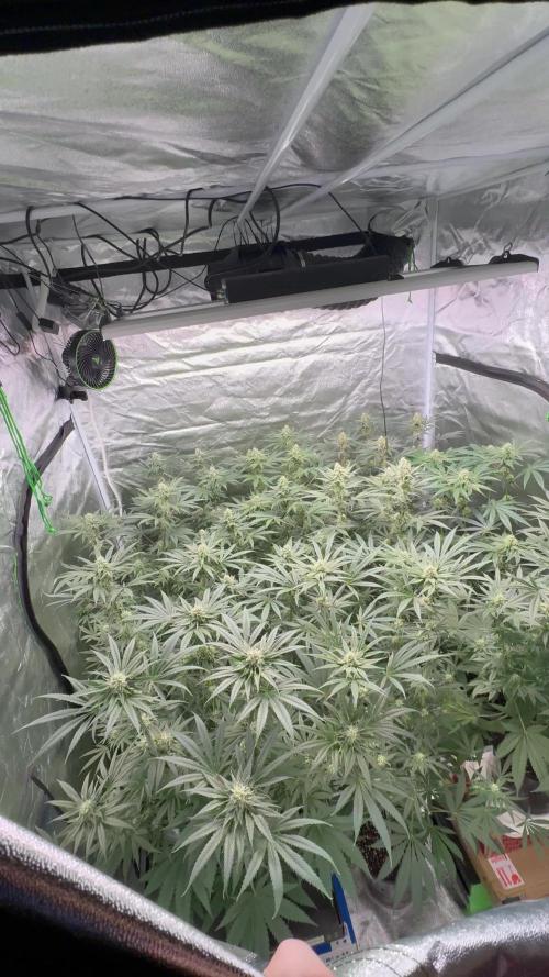
All the plants look great busy week check back next week for individual photos they should be getting fat by then

Likes
13
Share


@ChitownCannaChica
Follow
Plant is starting to look really nice and purple! Just been dunking the solo into water with feed to keep it moist! That’s the hardest part. Otherwise, just keeping her from falling over is the only thing! ✌️💚🌿💨
Likes
290
Share


@Ferenc
Follow
Beautiful.... a lot of LST made her like a snake. She would be very tall otherwise. Crazy...... a lot of rain by the way the temperature is getting low.... but it seems to be good all the things :)
I am not sure about her height may be even 100 cm over 🤪
Day 111: She looks like kind a strated the flowering cycle I mean the greenish tops I need to force her to flower shit weather UK it will be too late so I decided to do it manually covering her first 36 hours and then every day to get less than 12 hours light or smthg like that l. 🙄 So in case she started it helps her to go on if not then she will. I am sure she started 🙃

Likes
18
Share


@Manic
Follow
The budlets have fully developed and are now starting to fatten up. There hasn't been any deformed leafs or signs of nutes burn so the flush of distilled water from last week seemed to work so I'll b using 1/4 the the recommended dosage for there first feeding this week. Ap is really showing her purple color with the budlets and her sugar leafs. Blueberry is more potent smell so we will see what this week has in store.
Processing
Likes
3
Share


@Ogbangbang2981
Follow
Every week I watch my girls grow into beautiful budding trees only difference is now she in flowering I take her outside to absorb some of the beautiful sunshine and she's loving every bit of it. I let her stay outside for 8 hours in nature then back inside for another 8 hours of led full spectrum and turn out my lights for another 8 hr she's an Auto so she loving every minute of this ...... this is the results ⬆️
Likes
5
Share


@Mr_nugs_lover_David
Follow
Amazingly beautiful pheno right here this hard diesel by gea seeds has a pretty strong smell super gassy and sweet and the flowrs are just stunning, super beautiful, compact and completely covered in thc glands, it's such a pleasure, Stay tuned until the end to see the final result with FLO Living soil blend and water!! 💛💚❤️🔝👨🌾


Likes
44
Share


@Cogollo_eu
Follow
**Encontrarás la traducción a español al final de la descripción**
From/Desde: 22/03/19 || To/Hasta: 28/03/19
From day/Desde día: 29 || To day/Hasta día: 35
You can find the Money Maker Diary here:
** Podéis encontrar el diario de las Money Maker aquí:**
https://growdiaries.woodroom.tel/diaries/25667-gorillamakingmoney-gorilla-vs-money-m
-----IMAGES & VIDEOS-----
Video 1:
Exactly the same video as 2 but accelerated +400% to fix it to 1 minute for instagram.
Video 2:
7 days (29~35) TimeLapse, 1 image each 60 secs @ 30 fps
Video 9:
An overview video not as awesome as it's music 😂💪
Video 10:
Myself working on the babies, lowering tops, topping & training.
Video 12:
Full working on the secondary Gorilla, twisting some branches, topping & training.
-----WEEK SUMMARY-----
Well, now i'm getting a bit scared as i adventured myself into Photoperiod, high training, full topping... in indoors at once. I feel that plants are going good, but i really don't know what am i doing, and if i'm doing well. Also i don't know how much time should i give them of vegetation stage, because i don't want to eternalize myself with this tent, so i'm planning to leave this upcoming 6th week and maybe the 7th of vegetation, but no more. Anyway they look pretty atm 😻. In video 10 the main-linning Gorilla suffered an accident as i grabbed the main left branch from a weak point and it has broken (sec 39 aprox.) so it has one less node than the right branch.
-----WATERING CALENDAR-----
24/03/19 - 1.000 ml with all week nutrients @ 0.9 E.C. PH5.8
*****ESPAÑOL*****
-----IMÁGENES & VÍDEOS-----
Vídeo 1:
TimeLapse, exactamente el mismo vídeo que el 2 pero acelerado a un poco más del 300% para ajustarlo a 1 minuto para Instagram.
Vídeo 2:
7 días (29~35) TimeLapse, 1 imagen cada 60 segundos @ 30 fps.
Vídeo 9:
Un vídeo general no tan increíble como la música de fondo 😂💪.
Video 10:
Trabajando en las pequeñas, rebajando las alturas, haciendo topping y entrenando.
Video 12:
Trabajando a pleno en la segunda Gorilla, girando algunas ramas, haciendo toppings y entrenándola.
-----SUMARIO SEMANAL-----
Bueno, ahora estoy empezando a estar asustado. Me he aventurado a probar con fotodependientes, entrenamiento fuerte y super topping, todo a la vez. Siento que las plantas van bien, pero realmente no se lo que estoy haciendo ni si lo estoy haciendo bien. También tengo dudas sobre cuanto tiempo más dejarlas en estado vegetativo ya que no me quiero eternizar con este armario, por lo que estoy planeando dejar esta sexta semana de crecimiento y quizás una séptima, pero no más. Aun así y con todo, las nenas se ven preciosas 😻. En el vídeo 10 sufro un accidente en la Gorila con main-linning ya que amarré el final del tallo principal izquierdo por un punto débil y este se partió (sec 39 aprox.), por lo que tiene un piso de topping de más en comparación con tallo derecho.
-----CALENDARIO DE RIEGO-----
24/03/19 - 1.000 ml con todos los nutrientes semanales @ 0.9 E.C. PH5.8

Likes
8
Share


@Fuzzel
Follow
The get finally get going.
Only water this week.
Topping #1 and #2 over 5th node on day 33.
I guess I was kinda curious and ate the cut tops after giving them a rinse. They tasted great, kinda sweet&sour, reminded me of grapes that are not fully ripened and green. Also a bit parsly.

